OSTEOPATHY MATTERS
Manual Medicine Related News, Views and Curios
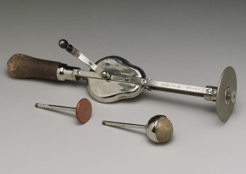
An account of a historical invention using mechanical vibration to treat pain

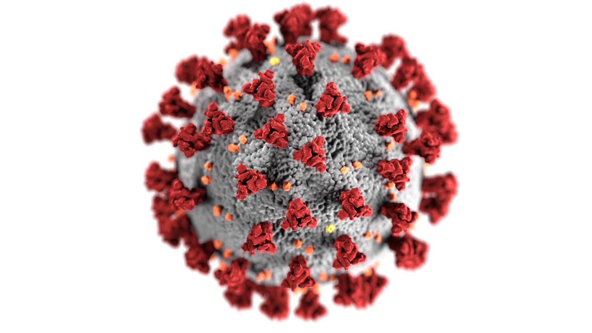
It seems that vitamin D may have a greater role to play in boosting our immune function than was previously thought. New research also indicates that a high dose Vitamin D supplement given to COVID 19 patients admitted to hospital may be as effective at reducing symptoms and preventing admission to intensive care as dexamathasone, cutting deaths by up to 60%. In addition, it is known that BAME communities in the UK often have low vitamin D status and this may in part be behind their greater apparent susceptability to COVID-19. Even before the pandemic many authorities had been suggesting that we should all take a vitamin D supplement during the autumn and winter months when sunshine is in short supply ( Spector Tim D, Levy Louis. Should healthy people take a vitamin D supplement in winter months? BMJ 2016; 355 :i6183 )
Following advice from both the Government and our professional body, as of January 2021, we remain open for patient visits and treatment during the latest Tier 5 lockdown, subject to the usual precautions. Please do feel free to contact us if you have any queries or concerns

Christmas is looming and ‘mix up Sunday’ for baking the traditional Christmas cake, and allowing it to mature, has already been and gone. Cinnamon features widely in both traditional spiced drinks and cakes and pastries. It has a distinctive festive taste but recent research has indicated an unexpected possible side benefit from regular consumption. A study carried out at Tel Aviv University appears to indicate that the beta protein tangles which accumulate in the brain, eventually leading to Alzheimer’s disease, are modified and untangled when afflicted mice are fed an extract of cinnamon bark 1,2 The cinnamon extract resulted in improved cognitive ability, when given to the afflicted mice, so much so that their performance was close to the unaffected control group. Finding a drug or medication which has the ability to remove or modify these tangles has been the ‘Holy Grail’ of Alzheimer’s research, if the research is transferable to human populations it will be truly remarkable that an ancient, and now everyday, spice might hold an answer to a distressing and otherwise untreatable condition. It might be interesting if a human population, which traditionally consumes large amounts of cinnamon as part of their normal year round diet, could be identified and shown subsequently to have a dramatically reduced incidence of Alzheimer’s. It’s probably far too early to raise hopes, and like many substances, excessive consumption of large amounts of cinnamon, e.g in capsules, may be harmful. (Some types of cinnamon also contain coumarin and other substances which can be toxic to the liver if consumed in large regular amounts). Used as it has been for thousands of years, in small amounts as a spice, it is safe. So we can all in the meantime enjoy our mulled wine and stollen, and raise a glass to the potential power of cinnamon. 1. Frydman-Marom A, Levin A, Farfara D et al . Orally Administrated Cinnamon Extract Reduces β-Amyloid Oligomerization and Corrects Cognitive Impairment in Alzheimer’s Disease Animal Models. PLoS ONE 2011 2. Saeideh Momtaz, Shokoufeh Hassani, Fazlullah Khan, Mojtaba Ziaee, Mohammad Abdollahi , Cinnamon, a promising prospect towards Alzheimer’s disease, Pharmacological Research,Volume 130,2018, 241-258,
We all realise that these are very difficult times, and all of us are still adjusting to what is no longer a 'new' normal. It is frustrating and stressful but we have to do our best to keep going. I thought it important to re-assure you that, as healthcare professionals, we are permitted to remain open for face to face visits by continuing to maintain the safeguards that we already have in place including PPE, regular cleansing and online and in person triage. It is important to emphasise that you should not attempt to book an appointment if you have been advised to self-isolate or if you are suffering from any symptoms suggestive of COVID, including for example, loss of taste or smell, new cough or breathlessness, or any chills, temperature or fever. If you do recognise that you are suffering any of the symptoms above, or are unsure about your health please do call 111 or visit https://www.nhs.uk/conditions/coronavirus-covid-19/ for more information and do stay at home in the meantime Hopefully we can all look forward to something more optimistic on the other side of lockdown. Thank you

The biopsychosocial model of healthcare is not a new concept, and many osteopaths will be familiar with the work of Wadell and Main and their 'yellow flags ', and the potential effect of adverse 'mood' on patient recovery and prognosis. This contrasts with the effects described by Norman Cousins in his classic account of the 'healing power of comedy' Indeed, as long ago as 1911 one of the original osteopathic pioneers: Louisa Burns, was keen to research the physiological relationship between the mind and body. Ms Burns carried out a number of experimental studies and published the results. An excerpt is shown below, which although very much of its time, still poses some interesting questions. Nearly a 100 years later in 2010 Davis et al published the results of a study looking at the effects of Botox injections on the emotional experience. Participants were given positive and negative video clips to view (rather than the 'gloomy words' used in Burns' day) Those subjects who had received Botox were more affected by the negative video and more likely therefore, to be a little 'gloomy' as a consequence. So, an inability to engage facial muscles fully in response to external stimuli may have a direct effect on mood and potentially also on well being. L ouisa Burns Effects of Gloomy Ideas The blood pressure, pulse, respiratory movements, reaction time and dynamometer tests were taken, then words from one of the “gloomy” lists were pronounced, and the subject asked to give a synonym or related word in answer. Fifty words are usually about as much as the average person wishes to endure in such a test The results of these experiments may be grouped as follows: Blood pressure decreased, sometimes by thirty or forty mm., but usually ten or fifteen mm. of mercury. Pulse decreased, with occasional irregularities. Respiratory movements become irregular, sometimes with frequent sighings. Reaction time increased, sometimes almost doubled, for gloomy words; the usual increase is about .5 sec. per word. Dynamometer tests show decrease of strength of both hands, but especially the right, during and after the pronunciation and replies of the “gloomy” list. The gloomy lists are about as follows: • dark • forlorn • worry • pity • sorrow • labor • falter • hard • dull • silent • dying • moody • ill • stupid • sickness • failure • weak • fatal • peevish • restless • weight • shroud • torn • sleepy • dark • grave • worry • silly • mean • weeping • hopeless • false • blue • aches • heavy • frozen • sad • weary • broken • tomb • alone • poor • sorry • decay • faded • old • grief • timid A few people who were subject to slight hypochondria were employed as subjects. The gloomy list had not the least effect upon their physiological activities. Apparently the gloomy trend of thought is usual with them. Louisa Burns, M.S., D.O., D.Sc.O. 1911 Studies in the Osteopathic Sciences: The Physiology of Consciousness: Volume 3
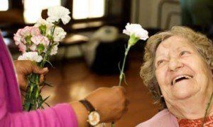
Kindness' as a taught objective does not feature prominently, if at all, in the curricula or course outlines of many osteopathic or medical institutions. This may seem anomalous, since as far as the recipient is concerned, kindness is one of the most notable qualities of an effective practitioner [. W G Pickering: Kindness, prescribed and natural, in medicine. J Med Ethics 1997;23:116-118]. We may be guilty of over emphasising the 'cold' scientific and quantitative elements of practitioner-ship at the expense of the 'warm' or more qualitative elements of human interaction. In some quarters there even appears to be a perception that 'kindness' is somehow a weak, non-scientific and archaic quality far removed, in clinical education, from the cold acquisition of specific academic and clinical skills. But, as practitioners, we are aware that kindness is a powerful factor and acts to not only enhance the therapeutic relationship, but also benefits the 'giver'. Indeed, Buddhist teaching has long advocated the merit of 'metta' a loving kindness and compassion. In addition, Professor Paul Gilbert has recently published 'The Compassionate Mind' which, from a western psychotherapeutic approach, reinforces the very same notion that kindness and compassion do seem to confer benefits on both the giver and the receiver. Clichéd as it may be, many of us feel that we have a genuine vocation to help others and have long recognised that there is much more to clinical effectiveness than the mantra of evidence based practice alone would imply. To paraphrase John Launer: “I’m not a clever osteopath, but I am a kind one.”[2. John Launer: On kindness. Postgrad Med J 2008;84:671-672.]

There has been an assumption, which appears to have some face value, that disability from low back pain (LBP) may be greater as a result of inactivity. A recent article suggests a complex interrelationship between obesity, inactivity and chronic back pain. Previous reviews had suggested that there was only a weak link with acute LBP, but a demonstrable and significant correlation for chronic LBP It may also seem self evident that a lifetime of relative inactivity could predispose to a greater risk of LBP. Waddell famously described LBP as a late 20th century epidemic and a healthcare enigma, and it shows no sign of letting up in the 21st century. Given our increasingly sedentary lifestyles, a number of researchers have tried to investigate a potential direct ink between weakness in the lumbar paraspinal muscles and increased risk of LBP. A Finnish study has demonstrated that, although obesity is still an independent risk factor for LBP, in obese subjects who are active the risk is reduced. A further study highlights the 'u' shaped realtionship between LBP, inactivity and excessive activity. To paraphrase Goldilocks it seems that the objective should be 'neither too much, nor too little' So, to safeguard their future, it would still seem prudent for all of us to continue to recommend structured physical activity and specific exercise for individuals with LBP when appropriate. .

A brief summary of the antimicrobial effects of copper and it's alloys.
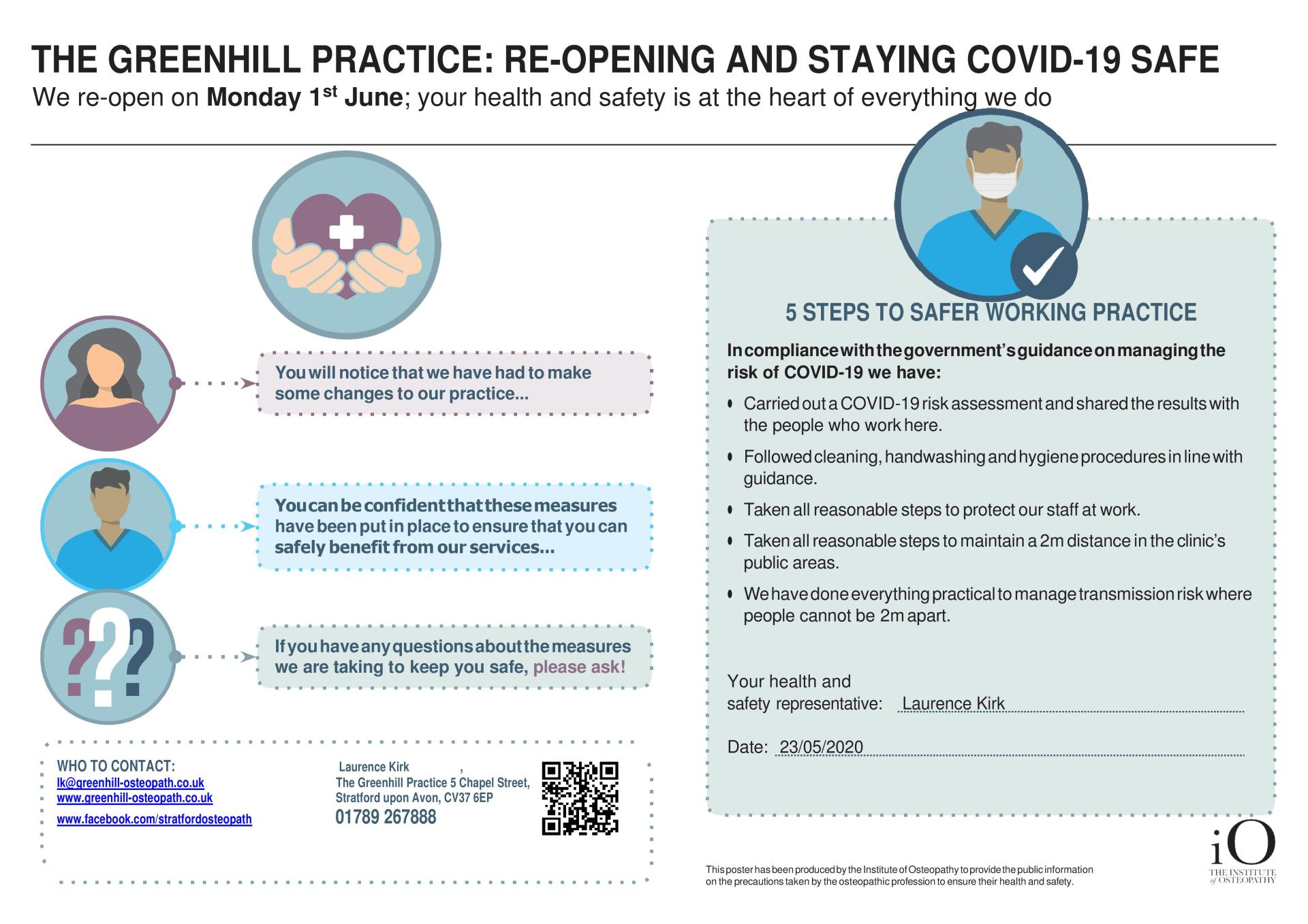
We are very pleased to announce that the clinic will re-open for face to face visits on Monday the 1st June following new advice from our professional body and insurers, now that the lock down measures have eased. Up until now physiotherapists, chiropractors and osteopaths were only advised to see 'emergency patients'; generally considered to be those with problems which would require emergency referral for swift medical intervention and couldn't otherwise be assessed remotely. Our insurance would potentially not cover us to treat in the circumstances as even seeing 'emergency' patients with the required PPE was considered to still be at the limit of acceptable risk. For that reason all but a small minority of local practitioners took the very hard decision, based on sound advice, to forego any income, and focus instead on keeping their patients, and the local community safe. Now that we have returned to practice we are obliged to work differently and screen for symptoms of Covid-19 prior to interacting with patients. We will do everything we can to maintain a safe environment, including wearing the required PPE with revised sanitising and cleaning regimes. All of your local practitioners are looking forward to seeing you once again and helping to relieve any pain or discomfort that you've been struggling with over the last few weeks.